


Introduction
Hyponatremic hypertensive syndrome (HHS) is characterized by hypertension, hyponatremia, and hypokalemia, which are attributed to unilateral stenosis or occlusion of the renal artery [1]. Approximately 16% of adults with unilateral renal artery stenosis have been reported to suffer from HHS [2]. The prevalence of HHS in children is not well known, but it is considered a rare disease [3,4]. However, a previous study reported that HHS was observed in 28% of pediatric patients with renovascular hypertension in a single center [2]. Patients with HHS may develop central nervous system symptoms such as convulsions, altered mental status due to hypertension, and combined hyponatremia [3,5]. Patients may also experience polyuria, polydipsia, and nephrotic-range proteinuria [2]. In most cases, the disease can be managed by addressing the underlying cause, controlling blood pressure, and providing supportive care for dehydration and hyponatremia [1]. Herein, we report a case of HHS in a 2-month-old infant who presented with nephrotic syndrome and improved after percutaneous balloon angioplasty.
Case report
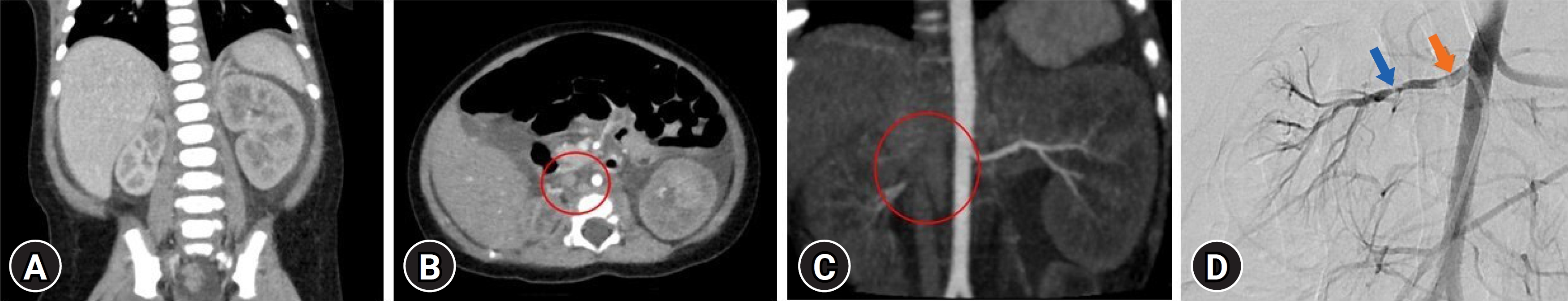
A 2-month-old male infant with no specific birth history presented to the hospital with general weakness, vomiting, and poor oral intake for 5 days. At the time of admission, the patientŌĆÖs initial body weight was 6.6 kg, which was 400 g lower than usual, and mottled skin was observed. His blood pressure was severely elevated, measuring 143/107 mmHg. Laboratory tests revealed proteinuria in the nephrotic range, with a urine protein-to-creatinine ratio of 107 mg/mg, as well as a low serum albumin level of 2.6 g/dL, low serum sodium level of 123 mmol/L, increased serum creatinine level of 0.8 mg/dL, and elevated renin and aldosterone levels of >80 ng/ml/hr (0ŌĆō3 years reference range: <16.6 ng/mL/hr) and 206 ng/dL (1ŌĆō11 months reference range: 6.5ŌĆō86.0 ng/dL). The patient developed atonic seizures and perioral cyanosis on the second day after admission. The patient received intravascular anticonvulsants, antihypertensive medications, and a 3% saline solution. Kidney Doppler ultrasonography revealed hypotrophy of the right kidney, decreased vascular flow due to narrowing of the right renal artery, and compensatory hypertrophy of the left kidney. Brain magnetic resonance imaging, electroencephalogram and tests for viral infections (toxoplasmosis, rubella, cytomegalovirus, herpes, human immunodeficiency virus, and syphilis) performed on this patient were all negative, and the next-generation sequencing panel for diagnosing podocytopathy-related gene mutations was negative. The patient had no significant family history related to thrombosis or stenosis. The patient underwent renal computed tomography angiography, and the results showed that the left renal artery was intact; however, the proximal ostium of the right renal artery was not clearly visible, and there was a suspected small right renal artery (Fig. 1A-C). The patient underwent percutaneous abdominal aortography and balloon angioplasty to improve right renal artery stenosis. Aortography revealed several focal stenoses in the right proximal and mid-renal arteries, suggesting thrombi accompanied by a focal filling defect (Fig. 1D). Through balloon angioplasty, the blood flow was improved by treating the stenosis. After the procedure, the patient's general condition improved, blood pressure decreased, and antihypertensive medication was tapered. After the procedure, serum albumin, sodium, renin, aldosterone, and creatinine levels were restored to normal. Proteinuria decreased after treatment, but nephrotic-range proteinuria persisted. There were no abnormal findings on antithrombin, protein C, protein S, antiphospholipid antibody, or factor 5, 7, 8, or 10 tests for detecting thrombophilia. Administration of low-molecular-weight heparin was initiated to manage the patient's residual renal artery thrombi, which was then switched to warfarin. Nineteen days after the procedure, the patient was discharged with warfarin and antihypertensive drugs. The proteinuria disappeared after 3 months without antiproteinuric medication. Six months later, a follow-up renal computed tomography angioplasty confirmed that the residual thrombosis had disappeared; therefore, warfarin was discontinued.
Discussion
Our case represents the first reported case of a neonate presenting with massive proteinuria, hypoalbuminemia, and hypertension caused by renal artery stenosis that was successfully treated with percutaneous renal angioplasty. These clinical features should be differentially diagnosed from congenital nephrotic syndrome.
Congenital nephrotic syndrome is a heterogeneous group of disorders characterized by nephrotic-range proteinuria and hypoalbuminemia, which typically manifests in infants younger than 3 months. It is a rare disease classified into primary congenital nephrotic syndrome, caused by genetic defects in podocyte proteins such as nephrin and podocin, and secondary congenital nephrotic syndrome, caused by infections (e.g., syphilis, toxoplasmosis, cytomegalovirus, rubella, hepatitis B, and human immunodeficiency virus) or maternal alloimmune disease (e.g., maternal systemic lupus erythematosus) [6]. Several case reports have demonstrated that HHS resulting from renal artery stenosis and thrombosis can lead to massive proteinuria and/or hypoalbuminemia [2,5,7,8]. Neonatal cases of nephrotic syndrome and HHS caused by renal arterial stenosis are rare. When ischemia occurs in the unilateral kidney, overstimulation of the renin-angiotensin-aldosterone system in the ischemic kidney causes hypertension in HHS. Additionally, hyperfiltration in the contralateral kidney leads to secondary polyuria, renal electrolyte loss, including natriuresis, and hypovolemia. The pathophysiology of massive proteinuria in HHS has not been clearly elucidated. Some theories include ultrafiltration, focal segmental glomerular sclerosis development, and podocyte injury associated with angiotensin II [2,9,10]. Therefore, patients with renal artery stenosis should be considered HHS due to renal artery hypertension. Renovascular hypertension in children is one of the most common causes of secondary hypertension. In previous studies, HHS was confirmed in about 1/4 of renovascular hypertension [2], so we should be careful not to underdiagnose HHS by reviewing the patient's clinical features and laboratory findings.
Patients diagnosed with HHS typically undergo treatment with antihypertensive medication and supportive care and may require invasive procedures and surgery to alleviate symptoms. Previously reported cases of neonates with HHS have been managed with medication and/or nephrectomy [4,5,11,12]. In our case, the patient underwent successful percutaneous balloon angioplasty, which preserved the function of the affected kidney. To our knowledge, this is the first report on using percutaneous balloon angioplasty to treat HHS in neonates. Percutaneous balloon angioplasty is an effective intervention for improving hypertension and correcting electrolyte imbalance in patients with HHS [2,8,13,14]. Angioplasty is considered the optimal choice for preserving kidney function in patients with renal artery stenosis, especially in children with a longer lifespan.
In our patient, multiple thrombi with renal artery stenosis were observed. The occurrence of thrombosis in neonates is rare. A German registry reported the incidence of symptomatic renal vein thrombosis in neonates to be at least 2.2 per 100,000 live births and renal artery stenosis is even more uncommon. This can be related to factors such as indwelling catheter insertion, for example, umbilical artery catheterization as well as disseminated intravascular coagulation, impaired liver function, fluctuations in cardiac output, and congenital heart diseases in neonates [5,15]. Our patient had no history of thrombosis, and thrombophilic conditions were excluded based on laboratory tests. However, it remains unclear whether renal artery thrombosis results in renal artery stenosis or whether hypoplastic kidney itself affects artery stenosis. However, renal artery stenosis and nephrotic syndrome can also cause thrombosis. Monitoring for additional thrombotic events is necessary in the future. At the last follow-up at 3 years of age, the patient did not experience any other thrombotic events.
Our patient presented with neurological symptoms at treatment initiation. HHS induces secondary hyperreninemia and hyperaldosteronism, further exacerbating salt and water loss and leading to malignant hypertension [2,4,13]. This condition has been associated with progressive target organ damage in children, including encephalopathy and intracranial hemorrhage. Central nervous system symptoms occur in approximately half of patients with HHS and are caused by encephalopathy resulting from severe hypertension and hyponatremia [2,3,5,7,8,12]. While most patients recover when electrolyte imbalance and hypertension improve, some patients may still have residual central nervous system sequelae [5,7]. Therefore, early detection and timely treatment of HHS are crucial for preventing neurological sequelae.
In our case, the presence of HHS provided clues regarding the underlying condition. Early diagnosis and appropriate management of HHS are essential to mitigate the risk of complications associated with persistent malignant hypertension and electrolyte imbalance in neonates.
 PDF Links
 PubReader
 ePub Link
 Full text via DOI
 Download Citation
 Print
Print