Introduction
Acute kidney injury (AKI) is a frequently encountered problem in premature infants, especially during the early neonatal period. In premature infant, one of the main pathogenesis of AKI is renal hypoperfusion secondary to cardiovascular compromise. Several perinatal conditions including perinatal distress and heart failure secondary to hemodynamically significant patent ductus arteriosus are common causes of AKI in premature infants [1].
Sacrococcygeal teratoma (SCT) is the most common neoplasm in neonates demonstrating an estimated incidence of 1 of 40,000 births and the complete surgical excision is the primary treatment of choice [2]. A subgroup of infants with huge and highly vascular SCT, however, frequently presents with fetal hydrops and high-output heart failure and is at high risk of mortality and renal morbidity. Several preoperative procedures that decrease the blood supply to the tumor and thereby stabilize the patientŌĆÖs hemodynamic condition and minimize intraoperative bleeding have been proposed. These include radiofrequency ablation, laparoscopic ligation and angiographic embolization of the feeding arteries [2,3]. Although a few cases of successful preoperative embolization using contrast media have been reported even in extremely premature infants [3┬Ł5], no detailed data have been reported regarding the short- and long-term outcomes of premature infants following radiological intervention using contrast media.
Here, I present the case of a premature infant with a huge SCT who survived preoperative embolization and mass excision but subsequently developed AKI that persisted until 4 months after birth. An overdose of contrast media was suspected as a significant contributor to the development of non-oliguric AKI.
Case report
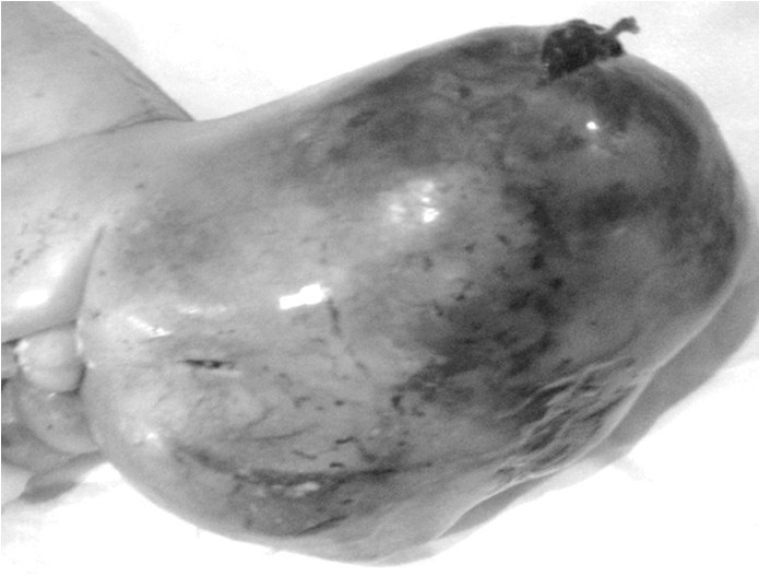
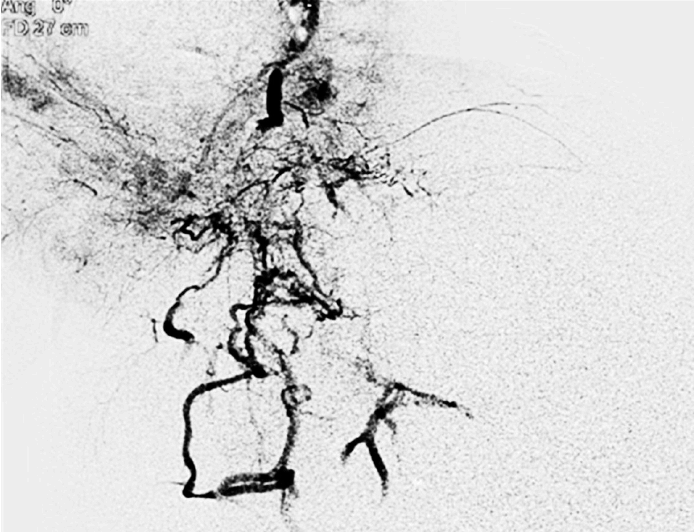
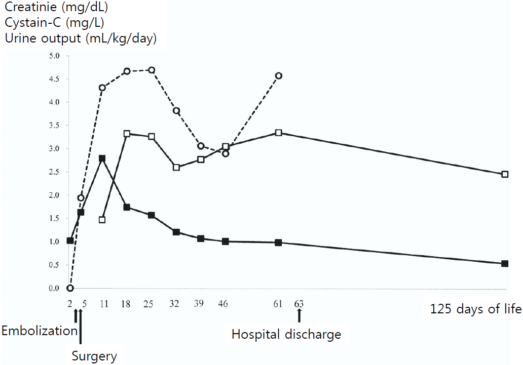
A large sacral mass was detected in a fetus by ultrasound at 23 weeks of gestation. The prenatal targeted ultrasound performed at 1 week later revealed a 96├Ś85 mm SCT with high vascularity that was invading the pelvic cavity. At 24 weeks and 3 days of gestation, ultrasound-guided laser ablation of the mass was attempted but failed. The size of the mass increased and severe polyhydramnios developed. The mother was hospitalized at 27 and 4 days of gestation because of premature rupture of membrane and chest discomfort. She was treated with antibiotics and a single course (2 doses) of antenatal betamethasone. At 28 weeks and 3 days of gestation, a boy was delivered via an elective cesarean section with a birth weight of 2,940 g including the huge pelvic mass (Fig. 1). The infant was intubated for respiratory distress in the delivery room and managed with positive pressure ventilation. The Apgar score was 3 and 5 at 1 minute and 5 minutes, respectively. On admission, there was massive bleeding from the ulceration site on the surface of the tumor and it was controlled by sutures. Severe anemia and the feature of disseminated intravascular coagulation were detected and corrected using blood products. Surfactant was also administered and ventilator care was required to maintain adequate ventilation and oxygenation. The infant was managed with several bolus infusions and inotropics, including dopamine and dobutamine due to systemic hypotension. On the second day of birth, abdominal and pelvic CT revealed a huge hypervascular mass with intrapelvic extension to the posterior aspect of the bladder. He was anuric for the first 2 days of life. On the third day of birth, embolization of the tumor was performed through the left common carotid artery. A 2.2-Fr probe was guided via the distal abdominal aorta to the bilateral internal iliac arteries and one lumbar artery. Occlusion of the feeding arteries was performed using gel forms, polyvinyl alcohol and metal coils. However, the intervention process was stopped because of the patient's unstable condition and the determination of a contrast media overdose (Iodixanol, Visopaque┬« 270, GE Healthcare, Cork, Island; a total of 25 mL was diluted with the same volume of saline). Post-embolization angiography still showed some remaining flow to the mass (Fig. 2). Immediately after the procedure, the patientŌĆÖs blood pressure increased and the dose of inotropics was tapered. He first voided at 12 hours after embolization. On the fourth day of birth, complete mass excision, including the pelvic portion of the mass, was performed. However, multiple blood transfusions were needed due to massive bleeding during the operation. He experienced cardiac arrest 3 times, presumably due to transfusion-associated hypocalcemia (lowest ionized calcium level: 0.20 mmol/L) and was resuscitated using intermittent cardiac compression (<1 minute for each duration) and multiple calcium infusions. The excised mass weighed 1,880 g (16.0├Ś11.0├Ś10.5 cm). Pathologic examination verified that the mass was a grade 3 immature teratoma. Urine output normalized on the day of the operation and thereafter, although urinary dribbling was observed. Neurogenic bladder was clinically suspected but urodynamic studies were not performed. However, neither the serum creatinine nor the cystatin-C levels normalized during the hospitalization period or by the time of the last follow-up examination (Fig. 3). The patient was weaned off the ventilator on 10th day of birth. No signs of hypoxicischemic brain injury were found on serial ultrasounds and brain magnetic resonance imaging performed at termequivalent age. He was discharged at 9 weeks of life (corrected age of 37 weeks and 4 days). Serial kidney ultrasound revealed persistent increases in the echogenecity of both kidneys, but they were of normal size without evidence of hydronephrosis. By 2 years of corrected age, the patient demonstrated rapid catch-up growth but development was significantly delayed for his corrected age. Neurogenic bladder persisted. There was no evidence of tumor recurrence on serial pelvic MRIs.
Discussion
Although the causes of prolonged AKI in our patient were not clear and might be multifactorial, the clinical course of our patient was suggestive of contrast-induced nephropathy (CIN) that resulted from an overdose of contrast media. CIN is generally defined as an increase in the serum creatinine level to >25% or 0.5 mg/dL over the baseline within 3 days following the administration of contrast media [6] and commonly manifests as a non-oliguric renal failure [7]. In this case, urine output was normalized following the surgery despite the prolonged decline in renal function as evidenced by the persistent increase in the serum creatinine level. Serum cystatin-C, another reliable marker of renal injury even in preterm infants [8], also did not normalize until the last follow-up examination. Among the many known risk factors of CIN, preexisting impairment of renal function is the most well-established [7]. In addition to immature kidneys with a low glomerular filtration rate, perinatal conditions that develop in newborn infants with huge SCT, including ŌĆ£steal phenomenonŌĆØ by the hypervascular tumor mass and unstable hemodynamic conditions, may contribute to the development of renal impairment. It seems unlikely that post-renal factors, the most common postoperative complications in the survivors of huge SCT, conferred the significant risk of development of AKI in our patient, because there was no apparent evidence of reflux or obstructive nephropathy on the follow-up examination.
The administered dose of contrast media is also a definite risk factor associated with the development of CIN. Although a large volume and multiple injections of contrast media have been reported in association with CIN in animal and human studies [9], no cut-off values have been established. The dose of the administered contrast media delivered to our patient (25 mL: approximately 8.5 mL/kg of body weight including the tumor) is similar to the suggested threshold of the total dose of a low-osmo larity contrast media (30 mL) that is associated with the markedly increased incidence of CIN in adults [10]. The manufacturer recommends that the intra-arterial dose of iodixanol administered to pediatric patients >1 year of age be 1-2 mL/kg, and the total dose should not exceed 4 mL/kg [11]. Although the epidemiology of CIN in pediatric pop ulations has not been well studied, the incidence of transient CIN within the recommended range has been reported as 10.6 % in pediatric patients who have received the intrave nous administration of the same agent [12]. In contrast, an other pediatric study on the contrast dose during cardiac catheterization reported extremely low adverse effects even when a large volume of a different kind of contrast media (Optiray 350 ┬«, Mallinkrodt, St Louis, Missouri, USA) was administered at a dose of Ōēź 6 cm3/kg [13].
It is still uncertain if even the complete occlusion of the feeding arteries could have reduced the risk of significant bleeding during the surgery because the chance of internal tumor bleeding and disseminated intravascular coagulation always exist, both prenatally and postnatally. A recent study reported the successful surgical resection of a giant SCT in a premature infant who was born at 30 weeks and 3 days of gestation following preoperative embolization [5]. Unfortunately, life-threatening cardiac arrest also could not be avoided, presumably due to hemolysis and transfusion-associated hyperkalemia. Our patient, who was born even more premature and with a greater tumor to birth weight ratio (0.63) than the previously reported case, also experienced a few life-threatening events during the operation. In recent 2 large-center studies, a survival rate of about 50 % was reported following surgical resection even in premature infants with large SCT who did not receive preoperative management including embolization [14,15]. A large body of evidence is needed to provide optimal treatment guidelines for giant SCT in neonates.
In conclusion, here I report the case of a premature infant with suspected CIN which was possibly related to an overdose of contrast media used for the preoperative embolization of a huge SCT. Further data on renal outcomes are needed to devise optimal dosing strategies for newborn infants receiving radiologic intervention, especially those at high risk of developing AKI.


 PDF Links
 PubReader
 ePub Link
 Full text via DOI
 Download Citation
 Print
Print


